Файл:Models of SMC and cohesin structure.svg
 Из Википедии, бесплатной энциклопедии

Размер этого PNG-превью для исходного SVG-файла: 234 × 598 пкс. Другие разрешения: 94 × 240 пкс | 188 × 480 пкс | 300 × 768 пкс | 401 × 1024 пкс | 801 × 2048 пкс | 512 × 1308 пкс.
Исходный файл (SVG-файл, номинально 512 × 1308 пкс, размер файла: 571 КБ)
Этот файл находится на Викискладе. Сведения о нём показаны ниже. Викисклад — централизованное хранилище для свободных файлов, используемых в проектах Викимедиа. |
Краткое описание
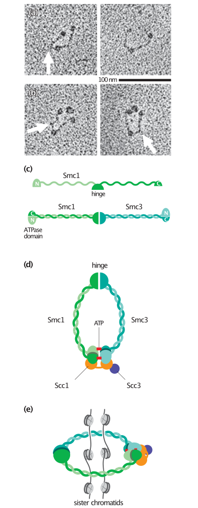
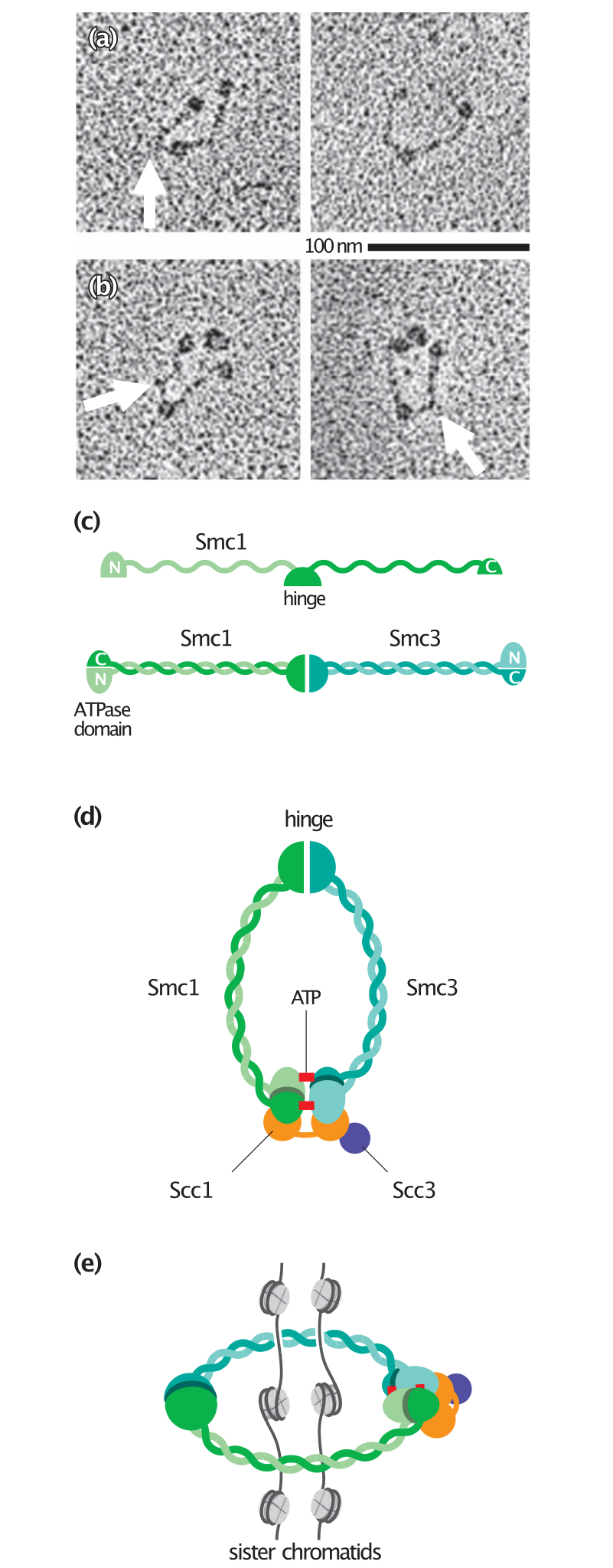
| Описание | English: a) Electron microscopy of Xenopus Smc1–Smc3 dimers illustrates the V-shaped structure commonly seen with SMC proteins. The flexible ‘hinge’ region is at the bottom of the V and the two globular ATPase domains are at the top. The arrow indicates a kink that is sometimes seen in one arm. (b) Addition of the two non-SMC subunits (Scc1 and Scc3) of the cohesin complex results in the appearance of a globular structure next to the two heads of the Smc1–Smc3 dimer. (c) The linear structure of an SMC protein includes two globular domains at each terminus, linked by a long repetitive sequence and a central dimerization or hinge domain. When the SMC protein is folded, the two domains at the termini join to form a complete ATPase domain, while the arm regions form a helical coiled-coil. The hinge domain that forms at the other end of the arm interacts with the hinge domain of another SMC protein. In cohesin, this results in the formation of a Smc1–Smc3 heterodimer. (d) Binding of ATP (red) promotes binding of the two ATPase domains, resulting in closure of the SMC ring. The non-SMC protein Scc1 interacts with both ATPase domains and holds them together. Cleavage of Scc1 in anaphase therefore opens the ring. (e) The cohesin complex may form a 50-nm ring around two sister chromatids. Because of its small size, however, this ring could only link nucleosomal DNA and not more complex chromatin structures. Panels (a) and (b) from Anderson, D.E. et al.: J. Cell Biol. 2002, 156:419–424.[1] |
| Дата | |
| Источник | The Cell Cycle. Principles of Control. |
| Автор | David O Morgan |
Лицензирование
 | Владелец авторских прав на этот файл разрешает всем использовать его в любых целях, при условии сохранения информации о владельце авторских прав. Разрешается распространение данного файла, создание производных произведений на его основе, а также коммерческое и любое другое использование. |
|
|
История файла
Нажмите на дату/время, чтобы посмотреть файл, который был загружен в тот момент.
| Дата/время | Миниатюра | Размеры | Участник | Примечание | |
|---|---|---|---|---|---|
| текущий | 22:18, 6 мая 2020 | 512 × 1308 (571 КБ) | Rob Hurt | Uploaded a work by David O Morgan from The Cell Cycle. Principles of Control. with UploadWizard |
Использование файла
Глобальное использование файла
Данный файл используется в следующих вики:
- Использование в en.wikipedia.org
- Использование в es.wikipedia.org